


The Heart of Excellence
Welcome to Cardiac Care Associates, where we're dedicated to your heart health with unwavering excellence. Your journey to a healthier heart starts here.
Experience Heartfelt Care with Cardiac Care Associates
Discover a new standard in cardiac care with Cardiac Care Associates. Our dedicated team of healthcare professionals brings together expertise, compassion, and innovation to deliver exceptional cardiac care services in Northern Virginia. We believe in treating more than just symptoms; we treat you as a person, understanding your unique needs and journey. Our commitment to excellence is evident in the personalized attention we offer every patient and our unwavering pursuit of the latest advancements in cardiac care.
Dive deeper into our story, our commitment, and see why Cardiac Care Associates is the trusted choice for heart health in Northern Virginia.

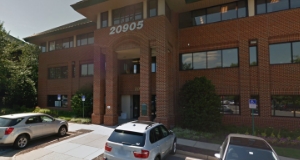
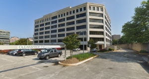
CARDIAC CARE LOCATIONS
.png)
.png)

.png)

.svg)
CARDIOLOGY SERVICES

Prevention, Care, and Management.
Tailored support for optimal well-being.
SERVICES COVERAGE:
.png)
Diagnostic Services.
Our cutting-edge imaging technology ensures accurate and comprehensive cardiac evaluations.
SERVICES COVERAGE:
.png)
Treatments and Therapies.
Discover a Range of Advanced Therapeutic Interventions for Comprehensive Cardiac Care.
SERVICES COVERAGE:
MEET THE DOCTORS


CARDIOVASCULAR DISEASE, NUCLEAR CARDIOLOGY, PERIPHERAL VASCULAR INTERPRETATION
Michael Banihashemi, MD, FACC, RPVI


.webp)

EXCELLENT PATIENT EXPERIENCE

“I have been visiting this practice on and off for over 20 years. The doctors and staff are phenomenal. They listen to you. Explain the information they develop. They provide you sound advice on improving your health. Staff are helpful. There are no long waits for your appointments. They are the best!”
— Verified Patient

“Quality organization staffed by the best cardiologists in Northern Virginia. Dr. B listens, assesses and provides his patients treatment options that are sound and achievable in a reasonable period of time. None better!”
— Verified Patient
“I have always had a great experience at Cardiac Care Associates with their excellent doctors and staff. Dr. Yager has consistently monitored my health and has always provided me the best consultation.”
— Verified Patient
“Dr. Elliot Mahlof is an excellent and caring cardiologist. Thanks to a recommendation from a friend who goes to Cardiac Care, my family found Dr. Mahlof and we are so grateful for his expertise, thoroughness, and attention to detail in caring for my daughter.”
— Verified Patient


LATEST NEWS & ARTICLES

Why Can I Feel My Heartbeat? Understanding Heart Flutters
Wondering "why can I feel my heartbeat?" Learn about heart palpitations, what causes that fluttering sensation, and when you should see a cardiologist.

Average BPM: What Your Heart Rate Says About Your Health
What is an average BPM? Learn what your resting heart rate says about your cardiovascular health, what factors influence it, and how to improve your numbers.

What is an Angiogram Procedure? A Complete Patient Guide
What is an angiogram procedure? Discover what to expect before, during, and after this important diagnostic test used to check for blocked arteries and heart disease.
.svg)
Secure Your Appointment Online Now!
To secure your appointment promptly, take advantage of our hassle-free online booking form.

Proud to be part of Privia Medical Group
Cardiac Care Associates is a proud member of Privia Medical Group. The best doctors in our community have joined together to form Privia Medical Group (PMG), a multi-specialty, high-performance medical group that puts patients first. Our physicians are united by the mission of providing better, more coordinated care for their patients.
To learn more about Privia Medical Group and find other Privia doctors, please visit our website.
Join Our Team: Exciting Career Opportunities Await!
Explore rewarding positions at Cardiac Care Associates. Join us in delivering top-tier cardiac care and making a positive impact on patient lives.
explore Open positions
Follow us on social media to stay updated with news and events!


